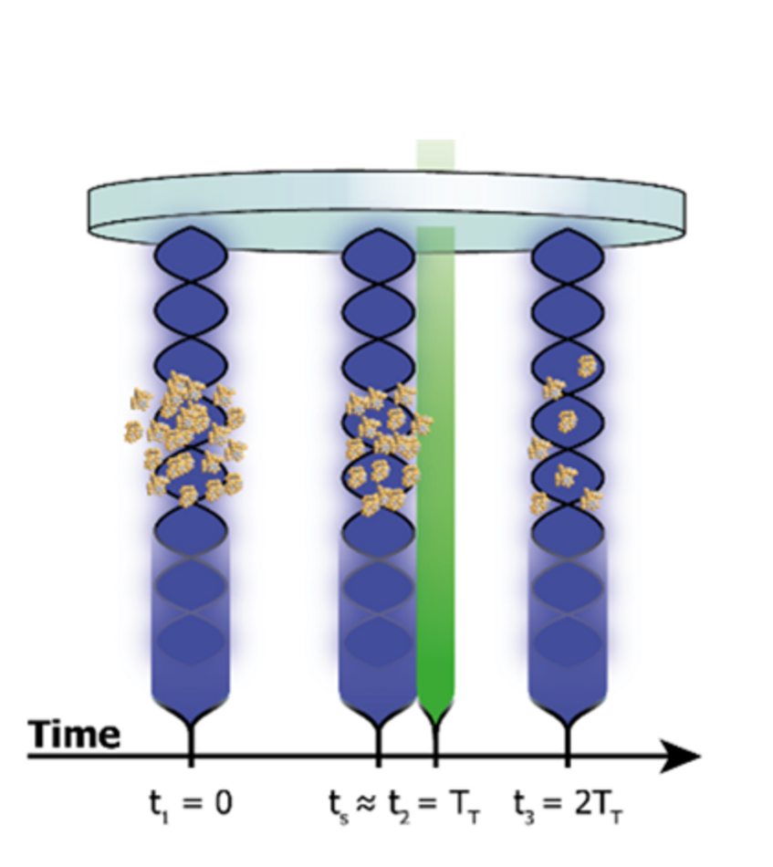
Figure 1:
Recoil spectroscopy in OTIMA interferometry: A laser (green) close to the second grating G2 can shift and blur the molecular interference pattern already even if every molecule absorbs less than a single photon on average [1,2].
Single photon recoil spectroscopy
Time-domain interferometry reduces the sensitivity to dispersive phase shifts and is therefore a promising tool for quantum-assisted molecule metrology. Here we present ideas how to measure the optical response of biomolecular systems using the recoil of zero, one or two photons in OTIMA interferometry [1].
The high sensitivitiy of the interference pattern to the recoil of a single optical photons can be used to measure absolute photoabsorption cross sections [2,3], by adding the running wave of an additional spectroscopy laser parallel to the grating vector of G2 (Figure 1). A molecule that absorbs a photon close to the second grating G2 receives a momentum kick and is deflected by Δx = nd2/λabs for the n-th Talbot order. A visbile photon (500 nm) will shift the first Talbot by about 1/6 of the grating period and cause a measurable reduction of the interference contrast.
Multi-photon recoil spectroscopy
Studying vibrational transitions requires photons in the near- or even far-infrared regime with wavelengths around 3-100 µm. The matter-wave fringe shift imparted by the recoil of a single photon can then only be resolved in high Talbot-orders, for slow molecules. A large shift can also be acquired by accumulating the recoil of many photons, but due to the anharmonicity of molecular states multi-photon-absorption is usually precluded. In complex molecules, however, vibrational excitations relax on the picosecond time scale, orders of magnitude faster than the pulse duration of a spectroscopy laser compatible with OTIMA interferometry. Therefore, multi-photon-absorption can lead to repetitive excitation followed by internal vibrational relaxation (IVR) and internal heating of the molecule.
To prevent internal heating, which might also cause spectroscopic shifts, we propose to use two photons with different wavelengths. The idea is illustrated in Figure 2a where two additional lasers L1 and L2 interact with the molecules close ot the Talbot-time TT. The absorption of a low-energy photon from L1 triggers the absorption of a photon of high momentum from L2, which then imparts the required kick and shift. Figure 2b sketches the transitions for double-resonant IR-UV-recoil spectroscopy. Since the IR absorption is required to enable the UV-absorption and kick, interference contrast will provide information about the absolute absorption cross section in the infrared regime.
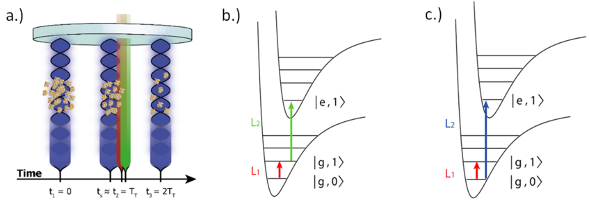
Figure 2:
a.) Timing of the five laser pulses for recoil-dip spectroscopy. b.) In double resonant IR-UV-recoil spectroscopy absorption of an IR photon in L1 enables absorption of the UV-photon in L2. c.) Depletion of the vibrational ground state [1].
Matter-wave-enhanced recoil dip spectroscopy
While the previous schemes exploited the loss of quantum interference to extract spectroscopic information, here we propose to restore and enhance interference contrast for spectroscopy. We exploit the resonant coupling of two lasers of different wavelengths to the same ground state |g,0>. High-energy photons of L2 can deliver a sufficient kick and reduce the matter-wave interference contrast. While the momentum transfer of L1 is insufficient to significantly influence the matter-wave fringe position, it can deplete the ground state that is shares with L2 . This suppresses the absorption of the more energetic photon and restores the interference contrast. Figure 2c shows the transitions at double resonant IR-UV-recoil spectroscopy.
Fluorescence recoil spectroscopy
If absorption is followed by fluorescence, the emission adds an additional recoil to the molecule and reduces the fringe contrast. This can be discriminated from the absorptive recoil, if the angle of incididence of the spectroscopy laser is rotated by 90 degrees. This can ensure that the absorptive recoil is along the grating lines or along the molecular beam and thus not perturbing the interference contrast. Figure 2a shows the operation principle.
Matter-wave-enhanced polarizability spectroscopy
The polarizability of a molecule provides information about the particle's composition, structure and van der Waals interaction with other molecules. In OTIMA interferometry we extract and rely on the optical polarizability at 157 nm to estbalish matter-wave interference, in the first place.
However, applying an additional tunable light grating G4 close to G2 (see figure 3) one can impose local dipole forces and an additional phase-grating. Tuning the frequency of G4 shall allow us to modulate the matter-wave fringe contrast and extracting the optical polarizability, even without the absorption of a single photon.
![]()
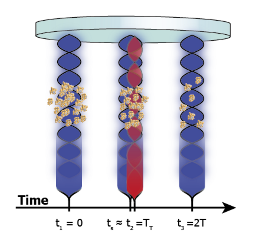
Figure 3: Polarizability spectroscopy with an additional fourth light grating close to the second grating of OTIMA.
References
- [1] Rodewald J., Haslinger P., Dörre N., Stickler B. A., Shayeghi A., Hornberger K., Arndt M.
New avenues for matter-wave-enhanced spectroscopy
Appl. Phys. B, 123:3 (2017). - [2] Eibenberger S., Cheng X., Cotter J. P., Arndt M.
Absolute absorption cross sections from photon recoil in a matter-wave interferometer
Phys. Rev. Lett. 112, 250402 (2014). - [3] S. Nimmrichter, K. Hornberger, H. Ulbricht, M. Arndt,
Absolute absorption spectroscopy based on molecule interferometry,
Phys. Rev. A 78, 063607 (2008).