Visualizing the Wave-Particle Duality with Single Molecule Resolution
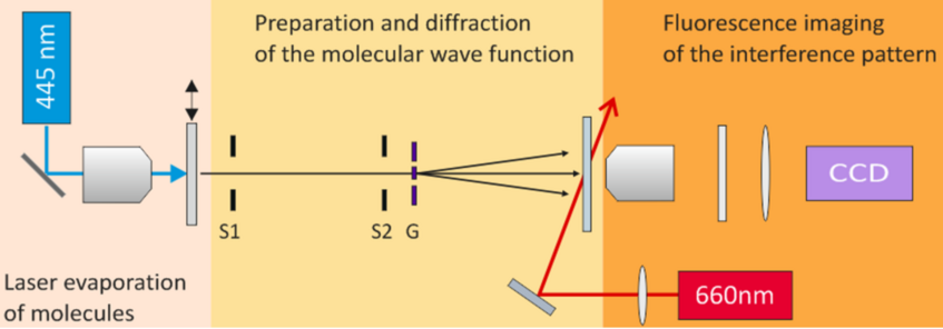
We launch a beam of molecules by focusing a 421 nm laser onto a thin molecular layer on the inner side of a vacuum window. The molecules absorb the radiation, are heated, and leave the surface with a typical velocity between 150 and 350 m/s. The strong confinement to 1.5 µm in the microfocus laser source prepares the molecular beam with a transverse coherence of around 5 µm at the grating, 1.5 m further downstream. The molecular beam is additionally laterally collimated to 10 µrad to prevent the diffraction orders from overlapping on the screen. Additionally, we select free-flight parabolas and thus the longitudinal coherence of the molecular beam. The nanomechanical masks were machined by our partners around Ori Cheshnovsky at Tel Aviv University. into ultra-thin membranes of different materials using a focused gallium ion beam.
About 60 cm behind the grating the molecules reach a 170 µm thick quartz plate, which is the vacuum-air interface. The molecules stick to the plate where they are imaged using laser-induced fluorescence. The fluorescent light is collected by a microscope objective and registered by an EMCCD camera. Collecting a large number of photons per molecule enables us to see every molecule individually and to determine their position with an accuracy down to 10 nm.
The video shows see single molecules arriving stochastically on the detector. As the experiment advances more molecules accumulate and the full deterministic interference pattern becomes visible. Although the position of each molecule in the pattern is random and cannot be determined before it actually arrives, the position where it may arrive is described strictly by diffraction.
- T. Juffmann, A. Milic, M. Müllernitsch, P. Asenbaum, A. Tsukernik, J. Tüxen, M. Major, O. Cheshnovski, M. Arndt, Real-time single-molecule imaging of quantum interference, Nat. Nanotechnol. 7 297-300 (2012)